Brachial Plexus Injury Diagnosis: 4 Key Tests Explained
Brachial plexus injury diagnosis is the critical first step toward effective treatment and recovery. Without an accurate diagnosis, the type and extent of nerve damage remains unknown and the right treatment cannot be planned.
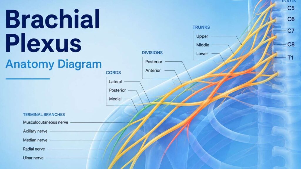
The brachial plexus is a complex network of nerves running from the spinal cord through the neck and into the arm. Injuries to these nerves produce symptoms that can mimic other conditions, which is why a structured, multi-step diagnostic process is essential.
This page explains how brachial plexus injury diagnosis works, what tests are involved, and why early evaluation by a specialist matters.
What Is Being Diagnosed?
Before understanding the tests, it helps to understand what doctors are looking for.
A brachial plexus injury can affect the nerves at different points along their path from the spinal cord (roots), through the neck (trunks and divisions), to the arm (cords and branches). The injury type determines treatment:
- Neurapraxia – nerve is stretched but intact; often recovers on its own
- Axonotmesis – internal nerve fibers are damaged but the outer sheath is preserved
- Neurotmesis – the nerve is completely torn or ruptured
- Avulsion – the nerve root is torn away from the spinal cord; the most severe type
Identifying where the injury is located (preganglionic vs. postganglionic) and how severe it is shapes the entire treatment approach whether conservative management, nerve repair, or nerve transfer surgery.
Step 1: Physical Examination
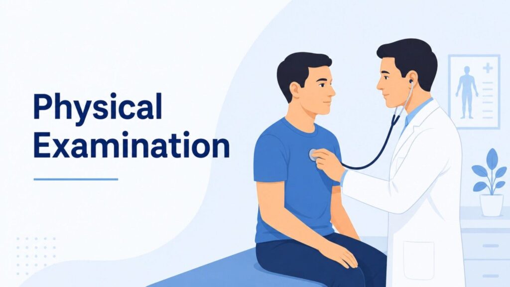
The diagnostic process always begins with a thorough physical examination.
During this assessment, the specialist evaluates:
- Muscle strength across the shoulder, elbow, wrist, and hand
- Sensation – testing light touch, pinprick, and temperature response
- Reflexes – biceps, triceps, and brachioradialis reflexes reveal which nerve roots are affected
- Range of motion in the shoulder and arm
- Horner’s syndrome signs – drooping eyelid, small pupil, and anhidrosis on one side of the face, which can indicate a C8–T1 root avulsion
A detailed physical examination can narrow down the level and likely severity of the injury before any imaging is ordered. It also establishes a baseline for tracking recovery over time.
Step 2: MRI Scan
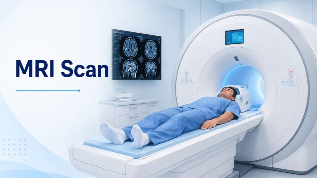
Magnetic Resonance Imaging (MRI) is one of the most valuable imaging tools in brachial plexus injury diagnosis.
What MRI reveals:
- Nerve root avulsions (pseudomeningoceles fluid pockets where roots have been torn from the cord)
- Damage to nerve trunks and cords
- Associated soft tissue injuries – muscle tears, vascular damage, haematomas
- Tumours or lesions compressing the nerve
High-resolution MRI of the cervical spine and brachial plexus is preferred. In some cases, MRI neurography a specialised protocol that highlights nerve tissue provides additional detail about the exact location and pattern of damage.
MRI is non-invasive and does not use radiation, making it a safe first-line imaging investigation for most patients.
Step 3: Electromyography (EMG)
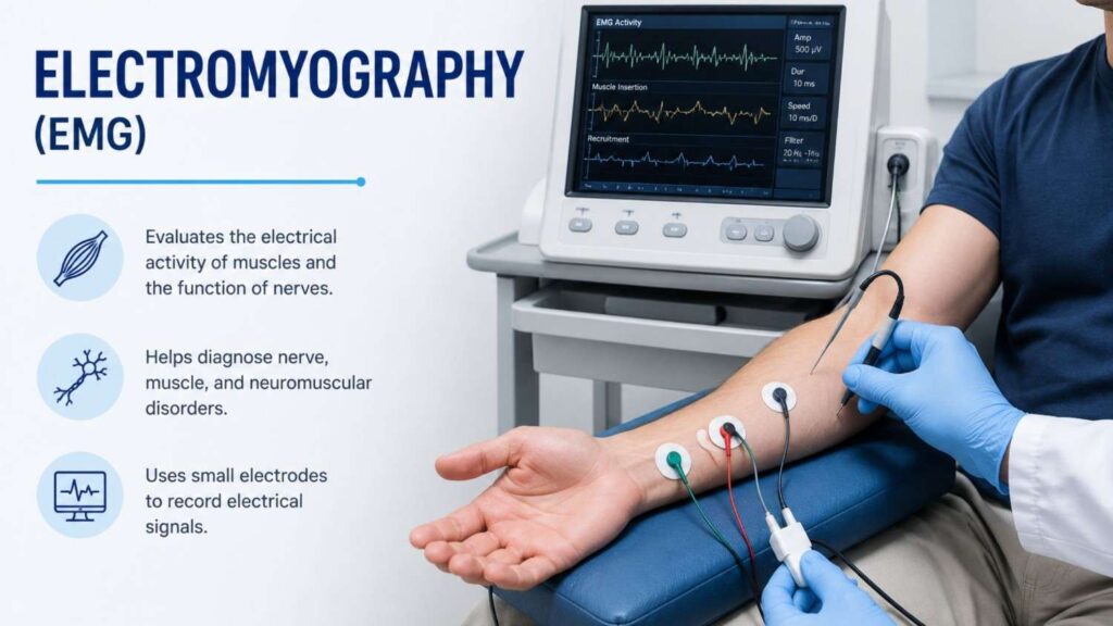
Electromyography, commonly called EMG, measures the electrical activity within muscles. It is a key component of brachial plexus injury diagnosis because it directly assesses how well the nerves are functioning.
During an EMG, fine needle electrodes are placed into specific muscles of the arm, shoulder, and hand. The electrical signals generated during rest and voluntary movement are recorded and analysed.
EMG findings in brachial plexus injuries:
- Fibrillation potentials and positive sharp waves – indicate active nerve damage or denervation
- Reduced or absent motor unit action potentials – suggest significant nerve disruption
- Polyphasic motor units – early signs of nerve recovery (reinnervation)
EMG is particularly useful for determining whether nerve regeneration is occurring which directly influences decisions about the timing of surgery.
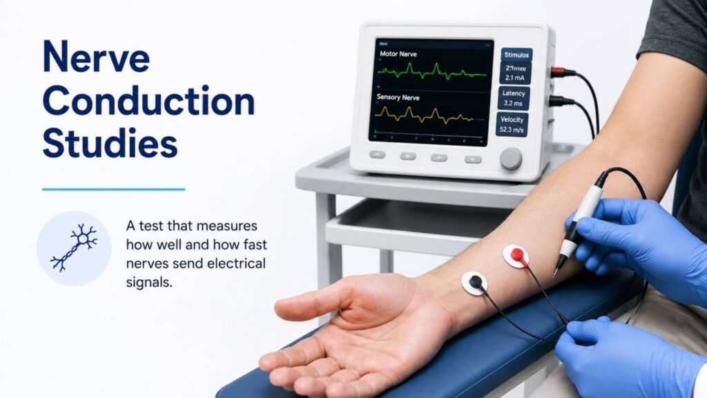
Step 4: Nerve Conduction Studies (NCS)
Nerve Conduction Studies are typically performed alongside EMG as part of a combined electrodiagnostic evaluation.
NCS measures the speed and strength of electrical signals travelling along a nerve. Sensors are placed on the skin over specific nerves, and small electrical impulses are delivered to stimulate the nerve at one point while recording the response at another.
Key NCS measurements:
- Conduction velocity — how fast the signal travels (slowed in injured or compressed nerves)
- Amplitude — the strength of the nerve signal (reduced in axonal injuries)
- Latency — the delay between stimulus and response
An important finding in brachial plexus root avulsions is a preserved sensory nerve action potential (SNAP) despite a clinically numb hand. This paradox occurs because the sensory cell body, located in the dorsal root ganglion, remains intact even when the root is torn from the spinal cord — a key marker that helps distinguish avulsion from other injury types.
When Should You Seek a Diagnosis?
Early brachial plexus injury diagnosis significantly improves treatment outcomes. Nerve surgery is most effective within the first three to six months of injury, before irreversible muscle wasting occurs.
You should seek specialist evaluation if you experience:
- Sudden arm weakness or inability to lift the arm after trauma
- Numbness, burning, or tingling from the shoulder to the hand
- Loss of grip strength
- Persistent shoulder pain following a fall, road accident, or sports injury
- Any arm symptoms in a newborn following a difficult delivery
The window for nerve reconstruction is time-sensitive. If these symptoms are present, early referral to a nerve specialist is strongly recommended.
Conclusion
A brachial plexus injury diagnosis is the foundation of every successful treatment outcome without knowing the exact location, type, and severity of nerve damage, no effective treatment plan can be built. Through a structured combination of physical examination, MRI imaging, EMG, and nerve conduction studies, specialists can map the injury precisely and determine whether conservative management or surgical intervention is the right path forward. Time plays a critical role in this process, as the window for nerve reconstruction surgery is limited and delays can permanently reduce the chances of restoring arm function. If you are experiencing arm weakness, numbness, loss of grip, or shoulder pain following trauma, seeking early specialist evaluation is the single most important step you can take.
For additional information about brachial plexus injuries and peripheral nerve disorders, patients can refer to the National Institute of Neurological Disorders and Stroke (NINDS).
FREQUENTLY ASKED QUESTIONS
Dr. Pradeep Kumar
Plastic & Reconstructive Surgeon – Brachial Plexus & Nerve Surgery Specialist
Dr. Pradeep Kumar is an experienced plastic and reconstructive surgeon known for his expertise in brachial plexus and peripheral nerve surgery. With extensive microsurgical experience, he focuses on restoring movement and function for patients with complex nerve injuries.
Sushrutha Institute of Plastic Surgery, Elite Mission Hospital, Thrissur