10 Powerful Ways HOW IS A BRACHIAL PLEXUS INJURY DIAGNOSED
A brachial plexus injury can affect the network of nerves that controls movement and sensation in the shoulder, arm, and hand. Understanding HOW IS A BRACHIAL PLEXUS INJURY DIAGNOSED is the first step toward receiving appropriate treatment and improving the chances of recovery. In Thrissur, early medical evaluation can help determine the severity of the injury and guide the most suitable treatment plan.
A proper diagnosis involves more than identifying pain or weakness. Healthcare professionals use physical examinations, imaging studies, nerve function tests, and medical history to understand the location and extent of nerve damage. Whether the injury results from trauma, sports accidents, childbirth, or other causes, accurate diagnosis is essential for restoring function and preventing long-term complications.
Why Early Diagnosis Matters
The brachial plexus is a complex group of nerves connecting the spinal cord to the shoulder, arm, and hand. Damage to these nerves can range from mild stretching to complete tearing.
Early diagnosis helps to:
- Identify the exact location of nerve damage
- Determine the severity of the injury
- Prevent permanent muscle weakness
- Improve recovery outcomes
- Plan appropriate treatment
- Decide whether surgery is necessary
Prompt diagnosis also helps distinguish a brachial plexus injury from fractures, tendon injuries, spinal cord conditions, or other neurological disorders.
Common Causes of Brachial Plexus Injury
Understanding the cause often provides important clues during diagnosis.
Common causes include:
- Road traffic accidents
- Motorcycle crashes
- Sports injuries
- Falls from height
- Industrial accidents
- Birth-related injuries in newborns
- Penetrating injuries
- Tumors affecting nerves
- Radiation-related nerve damage
Each cause may produce different injury patterns, making detailed evaluation essential.
Symptoms That Lead to Diagnosis
Many patients seek medical attention after noticing sudden changes in arm function.
Common symptoms include:
Severe Shoulder Pain
Pain often begins immediately after trauma and may spread into the arm.
Weakness
Patients may have difficulty lifting the arm or gripping objects.
Numbness
Loss of sensation may occur in specific parts of the shoulder, arm, or hand.
Tingling
Pins-and-needles sensations often indicate nerve involvement.
Loss of Reflexes
Reduced or absent reflexes may indicate nerve damage.
Muscle Paralysis
Severe injuries may cause complete loss of movement.
Burning Sensation
Some individuals experience persistent nerve pain.
Muscle Wasting
Delayed diagnosis may lead to shrinking muscles due to lack of nerve supply.
HOW IS A BRACHIAL PLEXUS INJURY DIAGNOSED Through Physical Examination
A detailed physical examination is the foundation of diagnosis.
During examination, healthcare professionals assess:
- Muscle strength
- Joint movement
- Arm positioning
- Grip strength
- Sensory changes
- Reflexes
- Range of motion
- Neck movement
- Shoulder stability
The pattern of weakness often helps identify which nerve roots are affected.
Medical History Plays an Important Role
Understanding how the injury occurred provides valuable information.
Questions may include:
- How did the injury happen?
- Was there a vehicle accident?
- Did the shoulder get stretched?
- When did symptoms begin?
- Is there severe pain?
- Has movement improved?
- Are symptoms getting worse?
- Any previous shoulder injuries?
The answers help narrow down the diagnosis.
Imaging Tests Used to Diagnose Brachial Plexus Injury
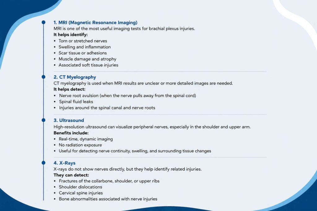
Imaging allows doctors to visualize nerves and surrounding structures.
MRI (Magnetic Resonance Imaging)
MRI is one of the most useful investigations.
It helps identify:
- Torn nerves
- Swollen nerves
- Scar tissue
- Muscle damage
- Soft tissue injuries
- Spinal cord abnormalities
MRI produces detailed images without radiation exposure.
CT Myelography
CT myelography may be recommended when MRI findings are unclear.
This test helps detect:
- Nerve root avulsion
- Spinal fluid leaks
- Damage near the spinal cord
It combines contrast dye with CT imaging.
Ultrasound
Ultrasound is increasingly used to examine superficial nerves.
Benefits include:
- Real-time imaging
- No radiation
- Dynamic assessment
- Cost-effective evaluation
It is especially useful for certain nerve injuries.
X-Rays
Although nerves cannot be seen on X-rays, this test helps detect:
- Broken bones
- Shoulder dislocation
- Cervical spine injuries
- Associated fractures
These injuries often occur alongside brachial plexus trauma.
HOW IS A BRACHIAL PLEXUS INJURY DIAGNOSED Using Nerve Tests
Nerve function testing provides important information about electrical activity.
Electromyography (EMG)
EMG evaluates muscle activity.
During the test:
- Small needles are inserted into muscles.
- Electrical signals are recorded.
- Muscle function is analyzed.
EMG helps determine:
- Which muscles are affected
- Severity of nerve injury
- Recovery progress
- Surgical planning
Nerve Conduction Studies (NCS)
NCS measures how quickly electrical signals travel through nerves.
The test identifies:
- Slowed nerve signals
- Blocked nerve transmission
- Severity of nerve damage
- Recovery potential
EMG and NCS are commonly performed together.
Neurological Examination
A neurological examination evaluates:
- Sensory function
- Muscle coordination
- Reflexes
- Balance
- Neck function
- Hand dexterity
This helps rule out spinal cord injuries or other neurological disorders.
Different Types of Brachial Plexus Injuries Identified During Diagnosis
Diagnosis also determines the type of nerve injury.
Neuropraxia
- Mild stretching
- Temporary nerve dysfunction
- Usually heals naturally
Rupture
- Nerve tears away from surrounding tissues
- Often requires surgery
Avulsion
- Nerve root separates from the spinal cord
- Most severe injury
- Surgical reconstruction may be needed
Neuroma
- Scar tissue develops around injured nerves
- Can interfere with nerve signals
Grading the Severity of Injury
Diagnosis determines whether the injury is:
Mild
- Temporary weakness
- Normal recovery expected
Moderate
- Partial nerve damage
- Recovery may take months
Severe
- Torn nerves
- Muscle paralysis
- Surgical treatment often required
Conditions That Can Mimic a Brachial Plexus Injury
Diagnosis also excludes other disorders such as:
- Rotator cuff injuries
- Shoulder dislocation
- Cervical disc prolapse
- Stroke
- Peripheral neuropathy
- Carpal tunnel syndrome
- Spinal cord injuries
- Fractures
Correct diagnosis prevents inappropriate treatment.
When Should Diagnostic Tests Be Performed
Timing matters.
Immediately after injury:
- Physical examination
- X-rays
- MRI if required
Several weeks later:
- EMG
- Nerve conduction studies
Some nerve tests are more accurate after the initial swelling decreases.
What Happens After Diagnosis
Once the diagnosis is confirmed, treatment planning begins.
Options may include:
- Observation
- Pain management
- Physiotherapy
- Occupational therapy
- Splints
- Nerve repair surgery
- Nerve grafting
- Nerve transfer procedures
- Muscle transfer surgery
- Rehabilitation
The treatment depends on injury severity.
Benefits of Accurate Diagnosis
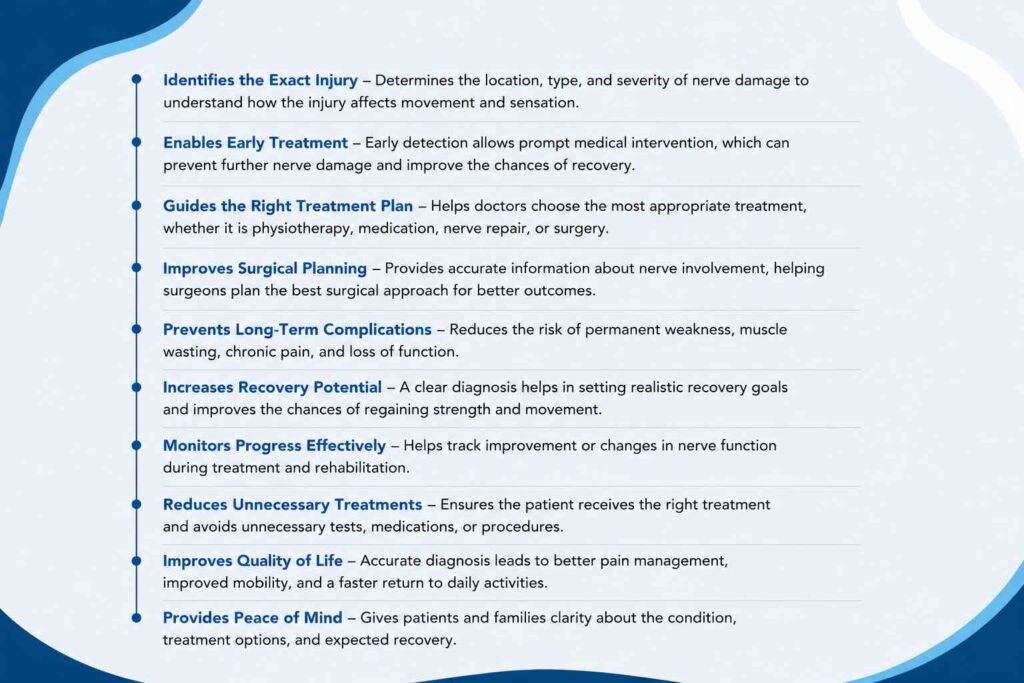
Proper diagnosis provides several advantages.
These include:
- Faster treatment decisions
- Better surgical planning
- Improved recovery chances
- Reduced complications
- Better pain management
- Clear rehabilitation goals
- Improved arm function
- Better long-term outcomes
Tips for Patients During the Diagnostic Process
Patients can support the diagnostic process by:
- Reporting symptoms accurately
- Mentioning previous injuries
- Attending follow-up appointments
- Completing recommended imaging
- Undergoing nerve studies
- Avoiding heavy lifting
- Beginning rehabilitation when advised
- Following medical instructions carefully
Recovery Depends on Early Diagnosis
Recovery varies based on:
- Type of injury
- Age
- Overall health
- Time before treatment
- Rehabilitation
- Surgical timing
- Extent of nerve damage
Some mild injuries recover within months, while severe injuries may require surgery and long-term rehabilitation.
Conclusion
Knowing HOW IS A BRACHIAL PLEXUS INJURY DIAGNOSED is essential for timely treatment and better recovery outcomes. A combination of physical examination, advanced imaging, and specialized nerve function tests provides a complete understanding of the injury. Early diagnosis allows healthcare professionals to develop an effective treatment plan, whether conservative management or surgery is required. If symptoms such as weakness, numbness, severe pain, or loss of arm movement occur after an injury, seeking prompt evaluation is the best step toward recovery. Patients in Thrissur can benefit from early assessment and a structured rehabilitation plan to maximize arm function and quality of life. The National Institute of Neurological Disorders and Stroke (NINDS) provides detailed information on brachial plexus injuries, including causes, diagnosis, and treatment options.
Frequently Asked Questions
1. How is a brachial plexus injury diagnosed?
2. Is an MRI necessary to diagnose a brachial plexus injury?
3. What is an EMG test for a brachial plexus injury?
4. Are nerve conduction studies painful?
5. Can X-rays diagnose a brachial plexus injury?
6. How soon should a brachial plexus injury be diagnosed?
7. Can a brachial plexus injury heal without surgery?
8. Why are multiple tests needed to diagnose a brachial plexus injury?
9. Can diagnostic tests determine if surgery is needed?
10. Does early diagnosis improve recovery from a brachial plexus injury?

Dr. Pradeep Kumar
Plastic & Reconstructive Surgeon – Brachial Plexus & Nerve Surgery Specialist
Dr. Pradeep Kumar is an experienced plastic and reconstructive surgeon known for his expertise in brachial plexus and peripheral nerve surgery. With extensive microsurgical experience, he focuses on restoring movement and function for patients with complex nerve injuries.
Sushrutha Institute of Plastic Surgery, Elite Mission Hospital, Thrissur